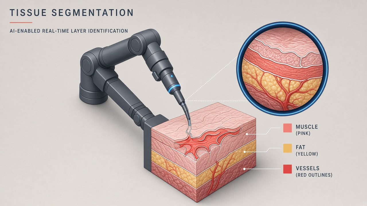
A surgeon operating the da Vinci robotic console sees magnified 3D video but must mentally infer where vessels, nerves, and fascial planes lie beneath tissue surfaces. Bleeding, smoke, and specular glare obscure boundaries within milliseconds. AI surgical robot segmentation applies deep learning to endoscopic frames, painting each pixel with a tissue class so software can highlight safe dissection planes, track instruments, or warn before critical structure contact. The research community has pursued this goal for over a decade through MICCAI EndoVis challenges; regulatory-cleared products for real-time autonomous tissue labeling inside the operative field remain rare compared with preoperative CT segmentation or post-hoc video analytics.
Robotics program directors, computer vision engineers, and regulatory affairs teams need a clear map of what works in benchmark datasets versus what Intuitive Surgical and competitors ship in cleared software today. Teams comparing assistive AI modalities can browse AI chatbot applications separately from SaMD-regulated segmentation. Additional medical AI explainers sit on the EliteAI.tools blog index.
What AI Surgical Robot Segmentation Means in Plain Language
AI surgical robot segmentation refers to machine learning models that classify each pixel in endoscopic or robotic camera video into anatomical categories such as instrument shaft, organ parenchyma, vessel, fat, or background, enabling software to understand tissue boundaries during minimally invasive procedures. Semantic segmentation assigns one label per pixel; instance segmentation additionally separates individual objects of the same class (two graspers, three vessel branches). Real-time inference requires models that run at 30 frames per second or faster on hardware mounted in or near the operating room.
Tissue boundary detection differs from instrument tracking alone. Early EndoVis challenges focused on robotic tool segmentation because tools have rigid geometry and high contrast against tissue. Labeling soft tissue is harder: color shifts with insufflation pressure, electrocautery smoke scatters light, and inter-patient anatomy varies. Modern pipelines fuse RGB video with depth or stereo disparity when available, following frameworks like SurgDepth that achieve state-of-the-art intersection-over-union on public robotic surgery datasets.
| Segmentation type | Output | Clinical use case |
|---|---|---|
| Semantic | Class per pixel (vessel, fat, tool) | Overlay navigation maps |
| Instance | Separate object masks | Instrument counting, collision avoidance |
| Binary boundary | Edge map between regions | Dissection plane guidance |
| Preoperative CT/MRI | 3D organ models | Planning, not live video (IRISeg class) |
da Vinci systems and the perception gap
Intuitive Surgical's da Vinci 5, FDA-cleared in March 2024, adds force feedback, upgraded 3D imaging, and Case Insights analytics, but cleared marketing materials describe video review and kinematic assessment rather than autonomous real-time tissue semantic segmentation during live surgery. Firefly fluorescence imaging highlights perfused tissue and bile ducts with injected dye, which is physics-based contrast rather than learned segmentation. The gap between research leaderboard scores on EndoVis datasets and cleared intraoperative AI overlays defines the current adoption frontier.
How the Underlying AI Pipeline Works
Typical surgical segmentation stacks capture endoscopic video, preprocess frames for color normalization, run a convolutional or transformer encoder-decoder network, and post-process masks with temporal smoothing across frames to reduce flicker. Training requires pixel-level annotations from expert surgeons, an expensive bottleneck that limits dataset size and induces domain shift when models trained on one hospital's lighting fail at another site.
EndoVis challenges and public benchmarks
The Endoscopic Vision (EndoVis) challenge series at MICCAI provides standardized robotic surgery datasets with instrument and tissue labels. EndoVis 2017 and 2018 established binary and multi-class instrument segmentation baselines; Surgical-DeSAM (2024) decouples Segment Anything Model (SAM) by replacing the image encoder with a Swin-DETR detector, achieving Dice scores of 89.62 and 90.70 on EndoVis 2017 and 2018 respectively. EndoVis 2022 SAR-RARP50 targeted semantic segmentation of six instrument classes plus ultrasound probe in robotic prostatectomy video. SurgDepth, published in 2024, fuses RGB and depth through Vision Transformer encoders and reached 0.86 IoU on EndoVis 2022, outperforming prior methods by at least four percentage points.
EndoVis 2024 and corruption robustness
The SegSTRONG-C sub-challenge at EndoVis 2024 tested instrument segmentation under realistic corruptions: artificial smoke, fake blood, low brightness, and background tissue changes. Data was collected on a da Vinci Research Kit (dVRK) platform at Johns Hopkins with replayed kinematics so identical trajectories could be filmed under clean and corrupted conditions. Top teams demonstrated that models trained only on pristine video degrade sharply when smoke or bleeding appears, a direct preview of domain shift failures in live operating rooms. Robustness-aware training with synthetic corruptions improves generalization but rarely closes the full gap to clean-set metrics.
Real-time inference architectures
Latency budgets drive architecture choices. Lightweight encoder-decoders (ENet, Fast-SCNN variants) and distilled transformers target 30+ FPS on GPU cards embedded in surgical towers. Surgical-DeSAM trades SAM's heavy image encoder for a detection-guided prompt path to approach real-time instrument masks. Temporal models (ConvLSTM, temporal attention) propagate labels across frames so a single misclassified frame does not flash incorrect boundaries to the surgeon. Multi-task heads jointly predict segmentation, depth, and tool pose to share features and reduce compute. Edge deployment on da Vinci compute modules requires validation against electromagnetic interference and sterilization-adjacent thermal limits in the OR.
Tissue boundary detection methods
Boundary-focused losses (Dice plus boundary F-score) penalize fuzzy edges along vessel walls and fascial planes. Active contour and level-set post-processing refine neural network outputs into smoother surfaces for augmented reality overlay. Stereo endoscopes supply disparity maps that disambiguate flat color regions where RGB-only models confuse fat and connective tissue. Some groups register preoperative CT segmentations to intraoperative video through deformable alignment, combining static 3D priors with live 2D predictions; registration drift when pneumoperitoneum shifts organs remains an open engineering problem.
Weakly supervised methods that train from bounding boxes or scribble annotations reduce surgeon labeling burden but produce coarser boundaries unsuitable for submillimeter vessel warnings. Self-supervised pretraining on unlabeled surgical video learns texture features that transfer to downstream segmentation with fewer annotated frames. Foundation models such as SAM require surgical-specific adaptation because natural-image priors misclassify specular highlights on moist tissue as distinct objects. The decoupled SAM approach in Surgical-DeSAM shows that detection-guided prompts outperform naive SAM fine-tuning on instrument masks while maintaining inference speed compatible with near-real-time display.
| Challenge / system | Reported metric | Deployment status |
|---|---|---|
| Surgical-DeSAM (EndoVis 17/18) | Dice ~90% instruments | Research benchmark |
| SurgDepth (EndoVis 2022) | IoU 0.86 semantic | Research benchmark |
| SegSTRONG-C (EndoVis 2024) | Robustness under smoke/blood | Challenge evaluation only |
| IRISeg 3.1 (Intuitive, K242461) | Kidney CT ML segmentation | FDA 510(k) cleared 2024 |
Typical development workflow steps
- Define target tissue classes and clinical scenario (prostatectomy, nephrectomy, hernia) with surgeon advisors.
- Collect de-identified video under IRB protocol; annotate frames with expert pixel labels or weak supervision.
- Train baseline UNet or transformer model; benchmark on held-out hospital splits to measure domain shift early.
- Augment with synthetic smoke, brightness jitter, and color grading to mimic OR variability.
- Optimize for latency on target hardware; add temporal smoothing and uncertainty highlighting.
- Run phantom and cadaver studies before any live human use under investigational device exemption if required.
- Prepare FDA premarket submission with software documentation, clinical validation, and cybersecurity per 2024 guidance.
Real Deployments and Published Evidence
Most published evidence for robotic tissue segmentation comes from MICCAI challenges and academic hospital partnerships rather than multicenter randomized trials of AI-guided dissection. da Vinci 5's cleared feature set emphasizes force measurement (up to 43% less tissue force in preclinical studies per Intuitive disclosures), Case Insights video analytics, and computing headroom for future vision software. IRISeg 3.1, cleared via FDA 510(k) K242461 in August 2024, segments contrast-enhanced kidney CT volumes for preoperative planning and intraoperative display, with clinicians required to edit ML-generated masks before clinical use. IRISeg demonstrates the regulatory path for Intuitive ML segmentation but operates on static CT, not streaming endoscopic tissue classification.
Research groups at Johns Hopkins, Imperial College London, and TU Dresden publish open models on EndoVis data with reproducible Docker evaluation containers. NVIDIA and startup collaborators demo OR overlays at conferences, but peer-reviewed outcome data linking segmentation-assisted navigation to reduced complication rates remains limited. Fluorescence-guided surgery (Firefly, ICG) is clinically established for specific structures; learned RGB segmentation aims to generalize beyond dye-dependent workflows at the cost of higher false-positive risk under domain shift.
Domain shift as the central deployment barrier
Models trained on da Vinci video from one institution often fail when camera white balance, insuffilation CO2 temperature, or patient BMI changes laparoscopic appearance. SegSTRONG-C quantified performance drops under smoke and bleeding corruptions that occur routinely during electrocautery. Continuous learning systems that adapt online raise FDA Predetermined Change Control Plan (PCCP) requirements finalized in December 2024: manufacturers must pre-specify modification protocols, validation methods, and impact assessments before deploying adaptive algorithms. Locked models are easier to clear but cannot recover from unseen OR conditions without manual retraining and new submissions.
Human factors and overlay design
Segmentation masks displayed as semi-transparent color washes must avoid obscuring blood color cues surgeons rely on to assess hemorrhage severity. Human factors guidelines from aviation and laparoscopic simulation recommend desaturated overlay palettes, adjustable opacity, and peripheral placement so the center of the field stays unobstructed. Audio alerts for high-risk structure proximity require low false-alarm rates; otherwise surgeons disable alerts within weeks. Usability studies under FDA guidance should measure task completion time, mental workload (NASA-TLX), and near-miss events with and without overlays in simulated bleeding scenarios before pivotal clinical trials.
Skill analytics products that segment instruments for postoperative review (Case Insights on da Vinci 5) occupy a lower-risk regulatory tier than real-time guidance because they do not influence decisions during tissue dissection. The segmentation research community should distinguish retrospective analytics from intraoperative decision support when citing EndoVis metrics to hospital procurement teams evaluating vendor claims.
Limits, Risks, and Ethical Guardrails
False-negative vessel segmentation can lull surgeons into unsafe dissection; false-positive boundaries cause unnecessary tissue sacrifice and prolonged operative time. Overlay latency above 100 milliseconds misaligns masks from moving tissue. Autonomy creep, where teams trust colored overlays over direct vision, is a documented human-factors risk in aviation-style safety analysis adapted to surgery.
- Annotation scarcity: Surgeon time for pixel labels caps dataset diversity.
- Generalization: Single-site training rarely transfers across hospitals without domain adaptation.
- Liability: Unclear whether overlay errors are device manufacturer or hospital responsibility.
- Cybersecurity: Networked vision modules in the OR expand attack surface per FDA 2024 software guidance.
- Equity: Training data underrepresents certain body habitus and skin tones inside the cavity.
Guardrails include surgeon-in-the-loop mandatory review, uncertainty heatmaps on low-confidence regions, kill switches that hide overlays instantly, and prospective registries tracking adverse events linked to AI assistance. IEC 62304 software lifecycle processes and ISO 14971 risk management apply to SaMD integrated with robotic platforms.
Who Should Use This and Who Should Wait
Academic robotics labs, MICCAI challenge participants, and hospitals with IRB-approved investigational studies should continue developing and validating segmentation models on public and proprietary datasets. Community hospitals should not deploy uncertified real-time overlays. Device manufacturers pursuing clearance should study IRISeg's preoperative CT pathway as a nearer-term regulatory milestone before live endoscopic tissue SaMD.
| Stakeholder | Recommendation | Guardrail |
|---|---|---|
| Robotics researcher | Benchmark on EndoVis; test corruptions | Report cross-hospital splits |
| Device manufacturer | Plan SaMD lifecycle per FDA 2024 AI guidance | PCCP for adaptive models |
| Hospital OR director | Use only cleared overlays; track outcomes | No investigational software without IDE |
| Surgeon end user | Treat overlays as adjunct to direct vision | Disable if latency or drift observed |
Frequently Asked Questions
Does da Vinci 5 perform real-time AI tissue segmentation?
FDA-cleared da Vinci 5 marketing describes upgraded imaging, force feedback, and Case Insights analytics, not autonomous real-time semantic tissue labeling on live endoscopic video. IRISeg covers CT-based kidney segmentation for planning.
Do EndoVis challenge winners work in real ORs?
Challenge metrics on curated datasets often overestimate clinical performance because test conditions exclude full domain shift from smoke, bleeding, and institutional camera settings. SegSTRONG-C explicitly measures corruption robustness gaps.
How does FDA regulate surgical AI segmentation?
Segmentation software that diagnoses, treats, or guides intervention is Software as a Medical Device requiring 510(k), De Novo, or PMA with clinical validation, cybersecurity, and software lifecycle documentation per 2024 FDA guidance. IRISeg (K242461) is a cleared Class II example for kidney CT.
Can domain shift be solved with more training data?
Multi-site datasets and corruption-aware augmentation reduce but rarely eliminate shift; prospective validation at each deployment site remains necessary. Adaptive models require Predetermined Change Control Plans for regulatory approval.
Is instrument segmentation clinically sufficient?
Instrument tracking supports collision avoidance and skill analytics but does not identify vessels or nerves beneath tissue surfaces. Tissue semantic segmentation addresses a harder and higher-risk perception problem.
Who is liable if an overlay mislabels a vessel?
Liability frameworks are evolving; surgeons retain primary responsibility for surgical decisions, while manufacturers carry device defect obligations under cleared intended use. Hospitals should define policies before adopting any assistive overlay.
How does Firefly fluorescence differ from AI segmentation?
Firefly uses near-infrared dye and camera filters to visualize perfusion or bile ducts, a physics-based contrast mechanism cleared for specific indications. Learned RGB segmentation attempts broader tissue classes without dye but with higher model uncertainty.
Open source, reproducibility, and clinical translation
EndoVis challenge organizers release training labels and Docker-based evaluation scripts so teams cannot tune hyperparameters on hidden test sets. Reproducibility accelerates translation when hospital research groups fine-tune public checkpoints on local de-identified video under IRB approval. Open weights without clinical validation still require full design control documentation if embedded in a commercial surgical platform. Partnerships between academic challenge winners and device OEMs remain the most common path from arXiv paper to pilot study, because OEMs own OR integration, sterilization workflows, and regulatory submissions.
Conclusion
AI surgical robot segmentation advances through EndoVis benchmarks, transformer and SAM-derived architectures, and robustness challenges that expose smoke and bleeding failures before they reach patients. da Vinci 5 expands sensing and analytics infrastructure while IRISeg clears a preoperative ML segmentation path for kidney CT. Real-time endoscopic tissue labeling inside the operative field remains largely investigational, constrained by domain shift, annotation cost, latency, and FDA SaMD expectations including 2024 Predetermined Change Control Plan guidance for adaptive models. Teams should treat leaderboard Dice scores as necessary but insufficient evidence, invest in multi-site corruption testing, and align product roadmaps with cleared use cases before promising autonomous tissue understanding in live robotic surgery.